Trauma des lateralen Kanthus (äußerer Lidwinkel)
Der äußere Lidwinkel vereint zwei besondere geometrische Merkmale: 1. die Position in Relation zum Augapfel, 2. der spitze Winkelgrad der Öffnung. Die seitlichen Lidbänder haben einen Ansatz innerhalb der Augenhöhle. Die Verletzung der Lidbänder führt nicht nur zur Lockerung der Ansatzes, sondern sekundär auch zur Deviation des Orbikularismuskels, der seine Verankerung im Lidwinkel verliert und sich wie ein Sphinkter nach medial bewegt. Dadurch verformt sich der Lidwinkel in einen stumpfen Winkel. Es ist schwierig die seitliche Verankerung des Muskels dauerhaft wiederherzustellen, da die chirurgische Fixation ständig der Muskelspannung ausgesetzt ist.
Abstract
The lateral canthus is the angle between the upper and the lower lid, where both are joined. The lateral angle has some principal characteristics like its position in relation to the globe and the degree of its aperture. The lateral canthal tendons attach the lids to the inner surface of the lateral orbital wall near the orbital rim. A lesion of the canthal tendons not only causes a laxity of the lids catnhal fixation, but also a deviation or the orbicularis muscle. If the insertion of the orbicularis muscle is lost at the canthal tendon, it can act like a sphincter and pull the lateral canthus towards the optical axis. It is dificult to maintain a permanent fixation against the traction of the orbicularis muscle. Therefore I imlant autologous fibrous tissue to replace the canthal tendons and create a permanent fixation.
 |
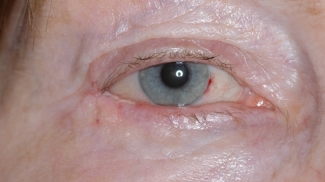 |
Durch die Läsion der seitlichen Verankerung entstand ein stumpfer Lidwinkel von 100°. |
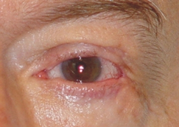
Durch die Rekonstruktion des lateralen Lidbandes wurde ein spitzer Lidwinkel von 30° wiederhergestellt und die Lidkante begradigt. |
 |
Runder lateraler Kanthus bei Zustand nach ausgedehnter Hundbissverletzung. Funktionelle Probleme der Lidmotilität ( Lidschlag und Lidschluss) |
|
Nach Wiederherstellung der lateralen Lidbandfunktion mit Peristolappen vom Jochbein. |
|
|